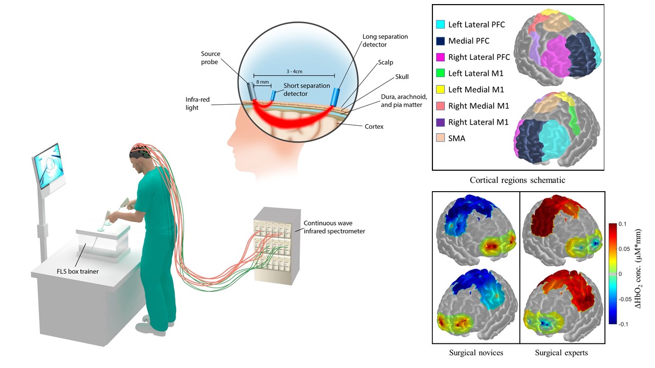
Measuring motor skill proficiency is critical for the certification of highly-skilled individuals in numerous fields. However, conventional measures use subjective metrics that often cannot distinguish between expertise levels. This is particularly evident in surgical training and assessment where current metrics are often subjective, rudimentary, and present inconsistent assessment conclusions. Our goal is to understand the dynamic mechanisms of motor skill learning, execution, and retention, within the context of robust and accurate motor skill assessment. We propose non-invasive brain imaging techniques, such as functional near-infrared spectroscopy (fNIRS), and electroencephalography (EEG) to measure changes in functional activation in real time for a variety of motor skill levels. Furthermore, we utilize machine learning concepts such as linear discriminant analysis, support vector machines, and kernel partial least squares, to precisely classify and predict motor skill levels that are independent of currently used subjective task performance metrics. This research has tremendous applications in surgical skill assessment, but also broader applications in stroke rehabilitation, and general motor skill theory.
Noninvasive Brain Imaging for Motor Skill Assessment
Motor skills that involve bimanual motor coordination are essential in performing numerous tasks ranging from simple daily activities to complex motor actions performed by highly skilled individuals. Hence, metrics to assess motor task performance are critical in numerous fields including neuropathology and neurological recovery, surgical training and certification, and athletic performance. In the vast majority of fields, however, current metrics are human-administered, subjective, and require significant personnel resources and time. Thus, there is critical need for more automated, analytical, and objective evaluation methods. We propose that fNIRS based imaging can accurately measure and classify subjects based on their expertise levels with significantly higher accuracy than currently established metrics in a prudent application: general surgery. By imaging regions critical to motor skill execution and learning, such as the prefrontal cortex, primary motor cortex, and supplementary motor area, we have shown that there are significant decreases in prefrontal activation and significant increases in primary motor area and supplementary motor area activation in surgical experts compared to surgical novices (shown below). For a more immediate impact, we also show evidence that metrics derived from brain imaging measurements are significantly more accurately in differentiating and classifying surgical physicians according to motor skill levels than currently used assessment metrics. These conclusions extend to medical students as they show evidence of decreased prefrontal activation and increased primary motor and supplementary motor area activation as motor proficiency is increased with training. Ultimately, our research has tremendous potential to accurately and robustly assess and predict motor skill levels that may present a paradigm change in technical motor skill assessment.

Noninvasive Brain Stimulation
The effect of transcranial electrical stimulation (tES) on human’s surgical motor skill learning behavior
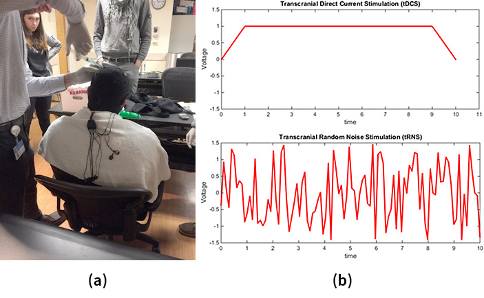
The application of transcranial electrical stimulation (tES) onto human’s brain has been demonstrated to modulate brain cortico-spinal excitability and cortical oscillatory patterns, as well as to induce significant modulation of behavioral performance in various domains including motor and visual ones, as well as executive functions involved in the learning processes. However, its effect on surgical tasks has not been studied yet. Transcranial direct current stimulation (tDCS) (Figure 1 (b) top) and transcranial random noise stimulation (tRNS) (Figure 1(b) bottom) are two promising methods in tES.
Our research is to study the effect of tDCS and tRNS on surgical motor skill learning, and also make comparison between these two methods, for future application of neuromodulation techniques on surgery trainees. The motor task we choose for the study is Peg Transfer designed by FLS (Fundamental of Laparoscopic Surgery) program. The task can effectively reflect human’s complex bimanual coordination and visuo-motor coordination ability. Two criteria are chosen to determine motor skill performance changes due to tES: standardized FLS score and the smoothness of the motion trajectory.


