Computational Biomedical Imaging

CeMSIM researchers are developing unique imaging modalities via the joint design of imaging hardware and image formation algorithms to provide new solutions with unprecedented capabilities for a wide array of biomedical applications. Researchers are active in integrating innovative imaging systems for precision medicine, pioneering quantum imaging and detection methodologies for biomedical applications, developing dedicated AI-based image formation/processing pipelines/diagnostic models and associated novel methodologies at the interface of AI and simulation-based inference.

Lead: Dr. Xavier Intes | Co-lead: Dr. Moussa Ngom
Projects

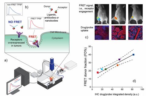
Breakthroughs in cancer biology have led to FDA-approved targeted therapies now standard in managing many malignancies. However, long-term, cancer-free survival remains a challenge, highlighting the need for further research on factors like drug-target engagement, a key to therapeutic success. In collaboration with Dr. Barroso's group at AMC, we have developed a fluorescence lifetime imaging technique to non-invasively quantify in vivo drug-target interactions in real time.

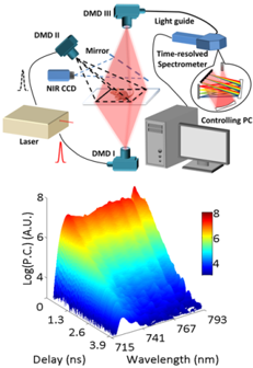
Unlike conventional imaging, which relies on a camera array, single-pixel imaging utilizes a single photodetector and advanced light modulation techniques to generate high-resolution 2D images from sequential measurements. At RPI, we have developed a groundbreaking imaging platform that integrates single-pixel imaging principles with spectral and nanosecond-scale temporal data acquisition. Enhanced by AI-driven processing, this innovative technique enables the rapid and simultaneous detection of multiple fluorescent species in biological tissues.

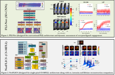
Over the past few decades, the design of biomedical optics systems, image formation, and analysis has been predominantly guided by classical physical modeling and signal processing approaches. More recently, deep learning (DL) has emerged as a transformative approach in computational modeling, offering significant advantages across diverse scientific fields and data analysis challenges.

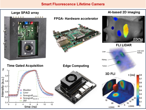
Hardware-based neural network accelerators for onboard computational imaging offer immense potential to enhance the efficiency, speed, and quality of imaging processes directly within devices. These cutting-edge solutions are poised to significantly impact medical technology. At RPI, we are leading the co-design and implementation of innovative onboard neural network accelerators to develop the first smart fluorescence camera. This breakthrough technology will enable real-time assessment of novel drug therapies and enhance fluorescence-guided surgeries.

The field of quantum metrology explores the impact of quantum mechanics on measurement systems and develops innovative technologies that leverage non-classical effects for enhanced performance. This research plan aims to establish a highly collaborative quantum metrology laboratory to image, isolate, and interrogate the properties of biological systems with unprecedented spatial and temporal resolution. The primary focus will be on optical techniques, given their unique relevance to biological imaging, sensing, and stimulation.

Advances in optical wavefront shaping have positioned multimode fibers (MMFs) as promising alternatives to single-mode fibers for high-data-rate communication and minimally invasive medical procedures. However, the complex modal behavior of light in MMFs limits their potential. We have developed a novel, single-step approach to retrieve the transmission matrix of MMFs, enabling coherent light propagation and effective phase correction.

Back to top